Melanoma: Clinic, diagnosis and treatment
Medical editor: Dr. Pierre Schneider, Dermatologist, Saint-Louis Hospital, France.
Related topics
- Oncodermatology
Key messages:
- Melanoma is an aggressive skin cancer: when faced with a patient with risk factors, it is advisable to implement preventative measures (using photo-protection and limiting sun exposure).
- Early detection of melanoma limits the severity of the tumor and reduces the risk of mortality.
- Melanomas are characterized by their stage (I to IV), their Breslow depth index and their BRAF status (mutated or wild type).
- Diagnosis is made by clinical examination of the suspected lesion, using the ABCDE method.
- In the case of a suspicious lesion, the patient should be referred to a dermatologist as soon as possible, who will then decide whether a diagnostic excision is necessary.
- Brain metastases represent up to 75% of metastatic melanomas.
- Melanoma is a type of skin cancer that develops from melanocytes, the cells responsible for producing melanin, the pigment that gives skin its color. It is considered the most serious type of skin cancer because of its ability to spread rapidly to other parts of the body and to metastasize.
- Melanoma usually appears as a nevus (or 'mole'), which is a spot or lesion on the skin that can change in size, shape or color over time.
- The main risk factors for melanoma include excessive exposure to UV radiation (from the sun or artificial sources), family and personal history of melanoma, light phototype (fair skin, blond or red hair, blue or green eyes), atypical moles, a high number of moles and a history of frequent or severe sunburn.
- Treatment for melanoma depends on many factors, including the size, location, and extent of the tumor, as well as the patient's general health. Treatment options may include surgical removal of the tumor, radiotherapy, chemotherapy, immunotherapy and targeted therapy.
- Melanoma is characterized by abnormal growth and proliferation of melanocytes.
- This tumor growth is often due to genetic and epigenetic mutations that alter the regulatory mechanisms of cell growth and programmed cell death.
- The most common genetic alterations in melanoma are mutations in the CDKN2A, NRAS, BRAF and PTEN genes, which regulate cell growth and proliferation.
- Mutations in the BRAF gene are the most common, with a prevalence of approximately 50% of cutaneous melanomas.
- Tumor cells may also secrete growth factors and cytokines that promote their own growth and the formation of new blood vessels, which supply essential nutrients to the tumor. This can promote the progression of the tumor and its ability to spread to other parts of the body.
- Exposure to UV radiation is the main modifiable risk factor for melanoma. UV radiation can damage cells’ DNA and lead to genetic mutations that promote the growth and proliferation of melanocytes. Excessive exposure to UV radiation can also suppress the immune response and promote melanoma progression.
- A family history of melanoma is also an important risk factor. People who have a first-degree relative with melanoma have a higher risk of developing the disease.
- Light skin is another important risk factor. People with fair skin have less melanin to protect their skin from UV radiation, making them more susceptible to sun damage. People with many atypical moles or who have suffered severe or frequent sunburns are also at greater risk of developing melanoma.
- Certain skin conditions, such as congenital giant nevus, can increase the risk of developing melanoma.
General characteristics
- Appears after puberty, over the age of 15.
- 80% of melanomas appear on healthy skin and 20% develop on a pre-existing "mole": change in color, shape and size.
- Usually found on the legs in women, on the back in men.
Most common patterns3
Superficial spreading melanoma (S.S.M.)
- It is the most common melanoma (70-80%). It corresponds to the multiplication of cancer cells horizontally in the epidermis.
- This phase lasts several years.
Without excision at this horizontal stage, the growth of the cancer cells becomes vertical:
- On the surface: appearance of nodules at the level of the lesion.
- Deep down: damage to the dermis and hypodermis.
Nodular melanoma (N.M.)
- It accounts for about 20% of cases.
- Its development is rapid with the appearance of a domed nodule, more or less pigmented, often brown black, corresponding to a vertical growth of cancer cells from the start.
- Some of these melanomas are achromic, giving a fleshy bud appearance which delays diagnosis.
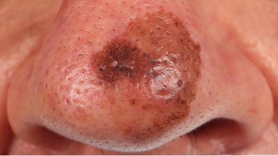
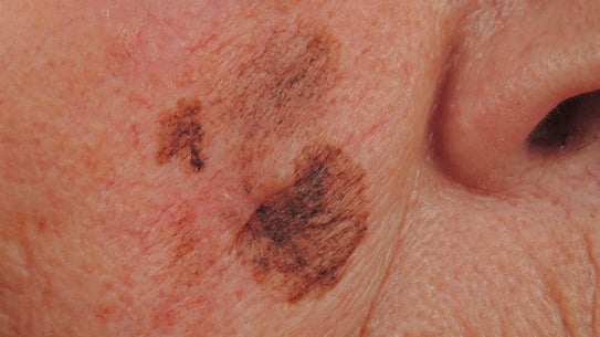
Dubreuilh's melanoma (L.L.M. = Lentigo Malignant Melanoma)
- It corresponds to 5 to 10% of melanomas.
- It affects elderly people in the face.
- It develops over a very long period of time, from 5 to 20 years.
- A pigmented, black or polychrome patch is found, with progressive extension.
- Late in the course of the disease, the appearance of a nodule, often greyish, signals the transition to the vertical phase.
Fig 1,2: Dubreuilh's melanoma
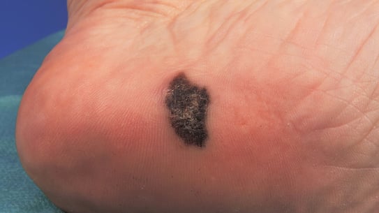
Acral Lentiginous melanoma (A.L.M.) or melanoma of the extremities
- It occurs on the palms of the hands, soles of the feet and under the nails.
- It occurs in phototypes with dark skin (V and VI).
Fig: Acral Lentiginous melanoma
Specific forms
Melanoma on pre-existing nevus
Nail melanoma
- Appearance of a longitudinal pigmented band, a few millimeters long, on a nail.
- Secondarily, the pigmentation extends to the skin at the base of the nail (Hutchinson's sign).
Mucosal melanoma
Poor prognosis
Melanoma in children
Although very rare, it poses the problem of early removal of extensive congenital pigmentary nevi.
Melanoma revealed by cutaneous, lymph node or visceral metastases
In these cases, the initial melanoma was either removed without histological examination (which is inadmissible) or regressed spontaneously in its entirety.
- Early diagnosis of melanoma is crucial for effective treatment and increased survival. Many actors can be involved in the early diagnosis of melanoma: GP or specialist, nurse, chiropodist, physiotherapist. Because of their position in the care pathway, GPs play a major role in screening people at risk and liaising with dermatologists1.
- The clinical examination is the first step in the diagnosis of melanoma. It consists of a thorough evaluation of the skin, looking for signs of melanoma such as suspicious pigmented lesions.
ABCDE method:
Clinically, the classic signs that should raise the suspicion of melanoma can be observed using the ABCDE method:
- A - Asymmetry: asymmetric lesion.
- B - Border: the edges of the mole are irregular, ragged or notched.
- C - Color: the color of the mole is not uniform. It may be shades of brown or black, or it may have patches of red or grey; it may also display two irregularly distributed colors.
- D - Diameter > 6 mm.
- E - Evolving: the mole is changing in size, shape, color or elevation.
- In addition to this ABCDE method, there is the Ugly Duckling method, which consists of screening for a malignant mole or spot by comparing it to others in the same area.
- In the presence of a suspicious lesion, the GP can assess of the risk level, and can then refer the patient to a dermatologist to screen for the presence of a melanoma. Only a biopsy (performed by a dermatologist) will allow the diagnosis of cutaneous melanoma to be reached by histopathology.
Factors to assess the level of risk1
- History of skin cancers (personal or family).
- History of sunburn with second-degree burns in childhood or adolescence.
- Numerous (≥ 40) or large (+ 5 mm) and irregular nevi.
- Exposure to artificial UV light.
- State of immunodepression, constitutional or acquired (immunosuppressive treatment, HIV).
- Occupational exposure to risk factors: UV, arsenic, polycyclic aromatic hydrocarbons (PAHs), ionizing radiation: people working outdoors, metal welding, iron and steel industry, medical and industrial radiology, or use of arsenical pesticides.
In the case of a patient with one or more risk factors and no suspicious lesions, the GP should encourage the patient to self-examine regularly (every 3 months) using the ABCDE method. It is important to clearly inform the patient about what should alert them:
- A melanocytic lesion that is different from his other moles already present on the body (Ugly Duckling method).
- A wound that does not heal.
- A mole that is getting bigger.
The treatment consists of several stages2:
First stage
Biopsy/exeresis for diagnostic purposes
The entire lesion is removed, without margin. This excision allows:
- Confirm the diagnosis of melanoma.
- To specify the signs of deep invasion in the dermis and hypodermis (Clark's invasion level).
- To measure, which is the main element, the Breslow index which corresponds to the thickness between the superficial cells of the epidermis and the deepest cancerous cell.
- To find the presence of ulceration on the surface of the melanoma, which plays a negative role in the diagnosis, and the presence of mitoses.
Second stage
Different situations may arise, depending on the Breslow index:
Diagnosis of a melanoma in situ
Strictly epidermal cancer cells: a surgical revision with a margin of 0.5 cm is done secondarily.
Diagnosis of a melanoma with a thickness (Breslow index) equal to or less than 1 mm
Surgical revision with a margin of 1 cm is necessary.
Good to know:
- The surgical revision consists of removing a somewhat wide band of healthy tissue around the scar from the first operation and going as far as the aponeurosis; this margin is called the safety margin.
- In the vast majority of cases, this surgical treatment allows patients to recover.
- Nevertheless, regular clinical monitoring, every six months for 3 years and then annually for life is necessary.
Melanomas with a thickness (Breslow index) of more than 1 mm
Surgically resected according to their thickness, i.e:
- Melanomas of 1 to 2 mm: surgical revision of 1 or 2 cm.
- Melanomas from 2 to 4 mm: surgical revision at 2 cm.
- Melanomas greater than 4 mm: surgical revision at 2 or 3 cm.
- Invasive Dubreuilh's melanomas require a 1 cm revision.
Patients are also subjected to a disease staging assessment with:
- Search for adenopathy:
- Clinical examination.
- Ultrasound.
- Possible search for sentinel lymph nodes. This is a specific examination, used in melanoma of the limbs to find the presence of micro-metastases at the level of the first lymph node relay of drainage.
- Search for visceral metastases:
- Thoracic, abdominal and pelvic CT, brain scan.
- Pet scan, especially for early pulmonary lesions.
- Patients are systematically referred to a multidisciplinary consultation meeting where the therapeutic solutions (adjuvant treatments) adapted to the patient and their melanoma are decided.
Good to know:
The recommended therapeutic approach depends on:
- The stage of the tumor (stages I to IV), determined according to the metastatic involvement (absent, locoregional or distant).
- For stages I and II (primary tumors without metastases): the Breslow index.
- The possibility of performing an excision or not (resectable or unresectable tumor).
- BRAF status of the tumor: BRAF mutated or wild type.
Advanced stages (III and IV) will systematically require immunotherapy as a first line of treatment. The choice of antibody will be determined by the BRAF status of the primary tumor. Adjuvant treatment or adjuvant surgery may also be given depending on the level of brain metastases, very common in metastatic melanoma (up to 75% of brain metastases) 4.
Prevention
During the summer
- No intentional exposure to the sun, especially between 12 noon and 4pm.
- Photoprotection by clothing is essential (long-sleeved T-shirt, wide-brimmed hat, sunglasses).
- Maximum sun protection* of exposed areas, twice a day, in the morning and early afternoon.
All other times of the year
- Maximum sun protection of exposed areas, twice a day, in the morning and early afternoon.
- Formally advise against the use of tanning booths (carcinogenic role of UVA).
* SPF ≥ 50 with the highest UVA protection guaranteeing a balanced UVB/UVA ratio (we now know the major role of UVA in the genesis of skin cancers, alongside UVB).
- Website of the Haute Autorité de la Santé (HAS, in French) : Mélanome cutané : la détection précoce est essentielle. [Website consulted on 27/02/2023].
- Website of the Vidal Campus (in French) : Mélanome cutané : recommandations de prise en charge. [Webstite consulted on 27/02/2023].
- Le mélanome de la peau : points clés - Mélanome de la peau (e-cancer.fr)
- Rajaonarison LA, Razafindrasata RS. Lésions cérébrales hémorragiques multiples révélatrices d’un mélanome métastatique [Multiple hemorragic brain lesions revealing metastatic melanoma]. Pan Afr Med J. 2019 Jun 3;33:78. French.
Create easily your professional account
I create my account-
Access exclusive business services unlimited
-
Access valuable features : audio listening & tools sharing with your patients
-
Access more than 150 product sheets, dedicated to professionals


